Hello everyone,
Here are the photos of the finished product. The main reason for waiting was the Weaver Eye Care Associates sign behind the reception desk, which you will see shortly. For now, some external shots:
Now for some interior shots after entering the vestibule, standing in the waiting area:
And now some pictures of the optical department:
And some pictures of the lighting that makes the optical look sooooo good:
Pictures of my office (spectacularly clean for the photos):
Looking at the insurance statements, and then FINALLY figuring it all out:
The pre-testing room, with an auto-lensometer (to read the prescription from your glasses) and an auto-refractor/auto-keratometer/corneal topographer that measures your refractive error and the surface of your cornea:
Some of my credentials, diplomas from the New England College of Optometry, Elizabethtown College and my Pennsylvania state optometry license:
Exciting pics ahead! Pictures of the exam room, complete with digital visual acuity chart (LCD monitor connected to the main exam room computer), exam chair and stand, slit lamp, BIO, manual keratometer, phoropter and handheld instruments. Main exam room computer is used for electronic medical records for a more modern look, keeping it 21st century:
Tile work in the bathroom and pictures of the lounge:
Pictures of the "second exam room," which is currently used as my specialized testing room. It has a lot of updated diagnostic equipment that allows me to manage and treat conditions such as glaucoma, macular degeneration and diabetic retinopathy:
On of the instruments in the diagnostic testing room is the HRTII, the Heidelberg Retina Tomograph II. This uses a scanning laser to analyze the optic nerve, which is essential in the diagnosis and management of glaucoma:
Opposite of the HRTII, is our digital retina photography setup and computerized visual field instrument.
A Topcon TRC-NW5 Polaroid camera was upgraded with a Canon Rebel digital camera that's connected to specialized software called Imacam. All pictures taken of the back of the eye (diabetic retinopathy, macular degeneration, choroidal nevi, glaucomatous optic nerves) are stored onto the computer and can be analyzed in detail by tools found with the program.
This compact computerized visual field unit is called the Oculus EasyField. It is used to measure a person's peripheral vision. It is helpful in glaucoma diagnosis, as well as measuring those with visual field loss from a stroke and those on high-risk medications such as Plaquenil that can affect the macula.
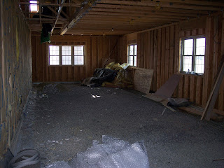
The total space available was 2500 sq.ft., which was a bit much for me as a start-up business. So the landlord allowed me to split the space down to 1250 sq.ft. This is the other unfinished half, which is pretty much what things looked like before the office even took shape. And then below is how the back hallway looked early on, and then the finished product:
And just a few more photos of the hallway, and optical/reception areas:
Hope you enjoyed the tour!
Take care,
Dr. Weaver


No comments:
Post a Comment